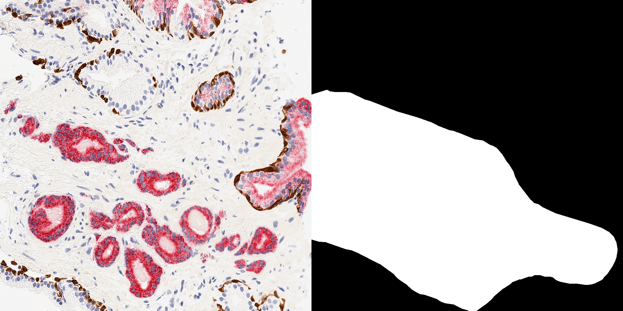
Cancer Cell Detection using Deep Convolutional U-Net
Overview
In this project, I developed a state-of-the-art deep learning model using PyTorch to automatically detect cancerous cells in tissue sample images. The model utilizes a deep convolutional U-Net architecture, which has shown remarkable performance in medical image segmentation tasks.
Motivation
Cancer diagnosis is a critical task in healthcare, often relying on manual examination of tissue samples by trained pathologists. However, this process can be time-consuming and subjective, leading to potential errors and delays in treatment. By leveraging advanced deep learning techniques, we aim to assist pathologists in accurately identifying cancerous cells, thereby improving diagnostic accuracy and efficiency.
Methodology
1. Data Preprocessing
- Data Splitting: Segregated the dataset into training and validation sets to monitor model performance and prevent overfitting.
- Image Resizing: Resized input images to a uniform size of 1024x1024 pixels to ensure consistency during training.
2. Model Architecture
- Deep Convolutional U-Net: Implemented a U-Net architecture consisting of encoder and decoder paths with skip connections, allowing the model to capture both local and global features effectively.
- Convolutional Layers: Utilized multiple convolutional layers with nonlinear ReLU activation functions to extract hierarchical features from input images.
- Pooling Layers: Employed max-pooling layers to downsample feature maps and increase the receptive field of the model.
- Transposed Convolutional Layers: Employed transposed convolutional layers to upsample feature maps and generate high-resolution segmentation masks.
- Skip Connections: Incorporated skip connections between corresponding encoder and decoder layers to preserve spatial information and facilitate precise localization of cancerous cells.
- Reference: The U-Net architecture used in this project was outlined in the paper "U-Net: Convolutional Networks for Biomedical Image Segmentation" by Olaf Ronneberger, Philipp Fischer, and Thomas Brox.
3. Training
- Dataset: Utilized a dataset comprising 1517 prelabeled images, with 175 images reserved for validation and 1342 images for training. Each image had dimensions of 1024x1024 pixels.
- Loss Function: Employed binary cross-entropy loss to optimize the model for accurate segmentation.
- Optimizer: Utilized Adam optimizer with a learning rate of 1e-4 for efficient model convergence and to avoid overfitting.
- Training Epochs: After 5 epochs of training, the model achieved an outstanding accuracy of 94%.
- Early Stopping: Analyzing the loss and accuracy trends revealed that approximately 3 epochs were sufficient to achieve full accuracy while avoiding overfitting.
4. Evaluation
- Metrics: Evaluated model performance using various metrics such as accuracy and loss to assess segmentation quality and convergence.
- Visual Inspection: Visualized model predictions overlaid on input images to assess segmentation accuracy qualitatively.
Results
The developed deep convolutional U-Net model achieved impressive results in detecting cancerous cells in tissue sample images. After 5 epochs of training, the model attained an outstanding accuracy of 94%. The dataset comprised 1517 prelabeled images, with 175 images allocated for validation and 1342 images for training. Each image had dimensions of 1024x1024 pixels.
The U-Net architecture outlined in the paper "U-Net: Convolutional Networks for Biomedical Image Segmentation" served as the foundation for the model development.
Upon analyzing the loss and accuracy trends, it was observed that approximately 3 epochs were sufficient to achieve full accuracy while mitigating overfitting, showcasing the efficiency and effectiveness of the model training process.
Future Directions
- Model Refinement: Continuously refine the model architecture and hyperparameters to further improve performance and generalization.
- Dataset Expansion: Incorporate additional annotated datasets to enhance model robustness and adaptability to diverse imaging conditions.
- Deployment: Explore opportunities to deploy the trained model in real-world clinical settings, potentially assisting pathologists in routine cancer diagnosis tasks.
Conclusion
In this project, I successfully developed a robust deep learning solution for automatic cancer cell detection in tissue sample images using PyTorch. By leveraging the power of deep convolutional U-Net architecture, the model demonstrated remarkable accuracy and holds great potential for enhancing diagnostic workflows in healthcare. This project exemplifies my expertise in deep learning, image processing, and their applications in solving real-world challenges, making me a valuable asset to any organization seeking innovative solutions in deep learning and image analysis.
This improves the accuracy and efficiency of cancer diagnosis, reducing errors and treatment delays.
Potential users are healthcare professionals, particularly pathologists, who will benefit from the automated detection of cancerous cells.
Python and Deep Learning were used to develop Cancer Detection U-Net.
